Research on the application of artificial intelligence in CT of digital oral cavity launched by Professor DING's team in Nature Communications
2022.05.06“When people set their mind on it, they can overcome anything.” Recently, Prof. Ding Zhongxiang (co-corresponding author), Department of Radiology, Affiliated Hangzhou First People's Hospital, Zhejiang University School of Medicine, cooperated with Prof. Shen Dinggang's IDEA Laboratory (School of Biomedical Engineering, Shanghai Tech University), "A fully Automatic AI system for Tooth and alveolar bone Segmentation Based on Cone-beam CT Images" was published in Nature Communications by several universities and hospitals with an impact factor of 14.919.
With the increasing awareness of oral health, digital oral technology has been widely used in the fields of orthodontic, prosthodontics, implantology, periodontal and maxillofacial surgery. Cone-beam CT (CBCT) images, with low radiation dose and high resolution digital images, can provide complete three-dimensional information of teeth and alveolar bone, and meet the needs of digital oral orthodontic functional alignment, implant surgery guidelines and various dental and periodontal surgery. However, the accuracy of the automatic segmentation algorithms is greatly challenged due to the fact that the quality of CBCT image is easily affected by artifacts, and there are great differences in dental morphology among different patients. To solve this problem, this study constructed a deep learning-based automatic tooth and alveolar bone segmentation system, which includes: 1) a hierarchical morphology-guided tooth segmentation network; 2) a alveolar bone filter-enhanced cascaded segmentation network.
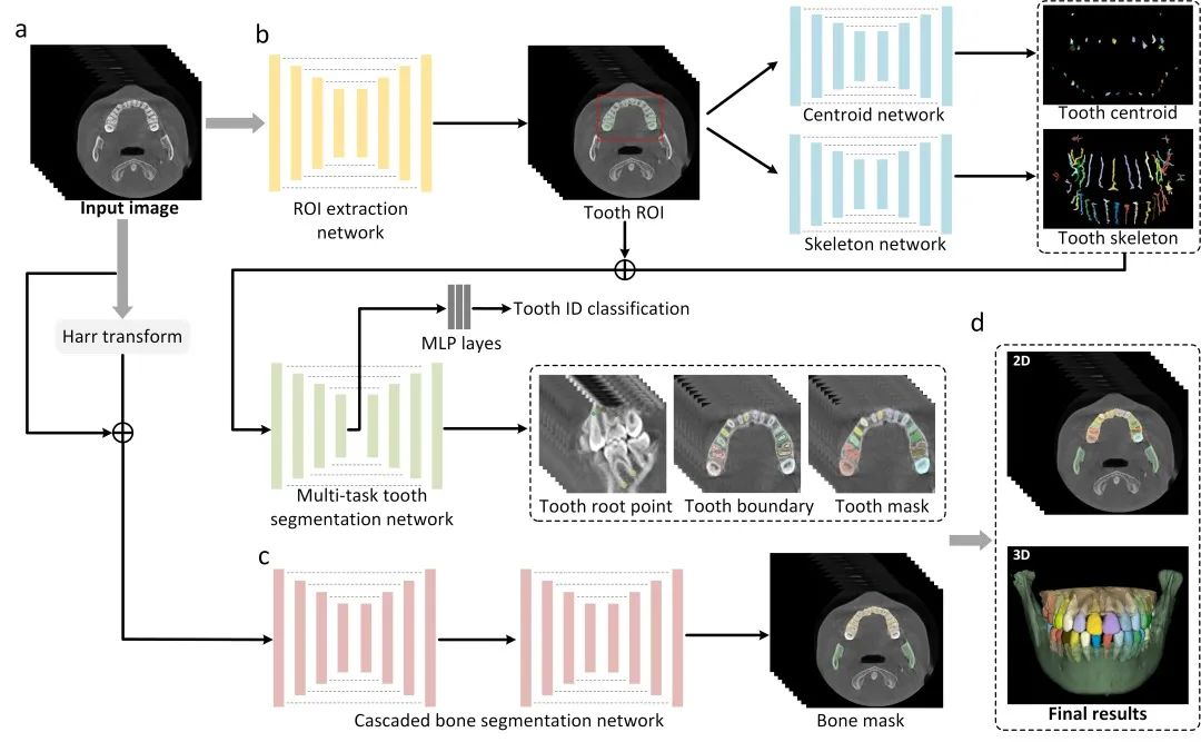
Fig. 1 Overview of AI system for segmenting individual teeth and alveolar bones from CBCT images. a) The input of the system is a 3D CBCT scan. b) The morphology-guided network is designed to segment individual teeth. c) The cascaded network is used to extract alveolar bones. d) The outputs of the model include the masks of individual teeth and alveolar bones.
In this paper, a hierarchical morphology-guided tooth segmentation network was proposed to accurately extract crown and root information of patients by characterizing "points, lines and planes" of tooth shapes from different angles for the first time, and constructed a high-precision 3-D tooth model for the digital oral cavity. In this study, 4,215 patients from 15 different centers (hospitals/dental clinics) obtained a total of 4,938 CBCT images, the largest international CBCT dataset to date. The research shows that the system not only greatly improves the clinical efficiency of digital oral cavity in the fields of orthodontics, prosthetics, implantology, periodontal and maxillofacial surgery, but also reflects that the combination of artificial intelligence and digital oral cavity will make a great contribution to oral medical services in the future.
Yu Bo, Prof. Ding's graduate student, is also one of the authors of this paper.